CAPILLARY RAREFACTION AND RIGHT VENTRICULAR DYSFUNCTION IN ADVANCED HFREF: HUMAN TISSUE HISTOMORPHOMETRY ANALYSIS
Introduction: Right ventricular dysfunction (RVD) limits survival in HF. Besides pulmonary hypertension (PH), load-independent mechanisms may contribute to RVD. In experimental PH, loss of myocardial capillary density (rarefaction) was implicated in decline of RV function.
Hypothesis: To elucidate the role of rarefaction in development of RVD in HFrEF.
Methods: We examined RV fractional area change (RV FAC%) prior heart Tx (median -57 days) in 124 consecutive HTx candidates (non-LVAD) and harvested RV free wall at HTx from patients in the bottom (Q1: 10-17%) and top (Q4: 32-42%) quartiles of pre-HTx RV FAC%. As controls (Con), we obtained RV samples from 20 unused heart donors. Quantitative histomorphometry and mRNA expression were studied.
Results: HF (65% non-ischemic) and Con were matched by age, gender and BMI. HF patients had larger and more impaired RV (RVD1 46 vs 32 mm, p<0.001, RV FAC 25±10%). Compared to Con, RV from HF displayed upregulation of PDK4 (Pyruvate Dehydrogenase Kinase4), NPPA (Natriuretic peptide A), BDH1 (β-OH-butyrate dehydrogenase), and downregulation of MYH6 (Myosin Heavy Chain 6).
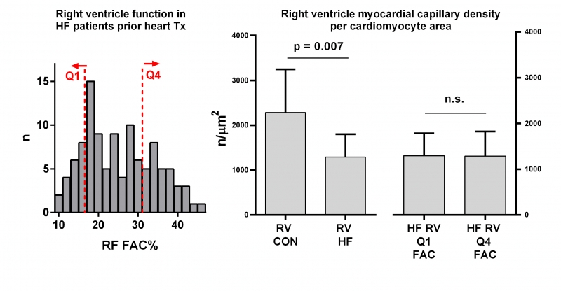
In HF vs Con, RV CM diameter was increased (25±6 vs 15±4 µm, p<0.001), but RV capillary density was lower (939±474 vs 1457±670 n/mm-2, p=0.007), even if adjusted to CM diameter (39 vs 102 mm-1, p=0.0005) or area (Fig). HF patients from Q1 and Q4 RV FAC differed by higher PH (mPA 37 vs 31 mmHg, p=0.01), larger RV size and higher PCWP (27 vs 22, p=0.002), but no difference in capillary density (unadjusted or CM diameter/area, Fig) or expression profile, HF etiology or comorbidities.
Conclusions: Compared to Con, RV of HF patients has reduced capillary density and myofilament remodeling. Yet, HF patients with lower RV FAC% do not differ from those with less impaired RV function. Capillary rarefaction per se does not explain RVD in HF.


