CONDUCTION SYSTEM PACING PRESERVES BOTH ELECTRICAL AND MECHANICAL INTERVENTRICULAR SYNCHRONY – A UHF-ECG VALIDATION STUDY
Background:
Interventricular mechanical delay (IVMD) is an established echocardiographic risk factor for pacing-induced cardiomyopathy development. Ultra-high-frequency ECG (UHF-ECG) is a non-invasive tool visualizing the ventricular activation sequence.
Aims:
To compare UHF-ECG interventricular dyssynchrony with echocardiography and to establish interventricular dyssynchrony related to conductive system pacing (CSP) and right ventricular pacing (RVP).
Methods:
54 patients with advanced AV conduction disease, without organic heart disease, and preserved LV systolic function were prospectively included. Thirty-three had RVP, and twenty-one had CSP. CSP included both His bundle pacing (n=5) and left bundle branch area pacing (n=16). UHF-ECG and echocardiography were obtained at the baseline and after 1 year of pacing. IVMD was manually calculated from standard echocardiographic projections. E-DYSV8-V1 was automatically calculated as a time difference between activation in V8 (LV free wall) and V1 electrode (RV free wall).
Results
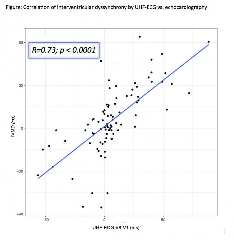
Both groups had similar baseline clinical characteristics and similar preimplant IVMD and e-DYSV8-V1. While interventricular dyssynchrony was not changed during CSP (mean change -0.37 ± 4.8ms, p=0.94 for IVMD and -1.7 ± 3.7ms, p=0.98 for e-DYSV8 -V1), it was significantly increased with RVP (mean change +29.3 ± 4.6 ms, p0.0001 for IVMD and +26.1 ± 5.1 ms, p=0.0001 for e-DYSV8-V1). There was a strong overall correlation between IVMD and e-DYSV8 -V1 in all studied ventricular rhythms (R=0.73, p=0.0001) - Figure.
Conclusions
UHF-ECG expresses the interventricular dyssynchrony noninvasively by measuring the activation difference between V8-V1 chest leads. RV myocardial pacing increases interventricular dyssynchrony, while CSP doesn’t.


