AUTOMATED DETECTION OF QS UNIPOLAR MORPHOLOGY AT ELECTROGRAPHIC FLOW MAPPING-IDENTIFIED SOURCES PRE- AND POST-ABLATION
Background
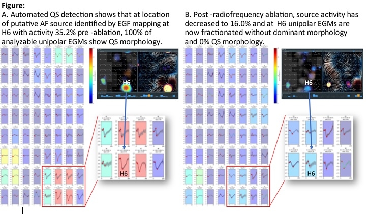
Electrographic flow (EGF) mapping estimates electrical wavefront propagation through atrial myocardium as a result of transmembrane voltage changes represented by unipolar electrograms (EGMs). Putative AF sources have been shown to manifest unipolar QS EGMs.
Objectives
Examine correlation between EGF-identified source locations and QS unipolar EGM morphology.
Methods
Recordings from 64-pole basket catheters in persistent AF patients were analyzed. For patients with a putative AF source identified by EGF and had source ablated, we analyzed unipolar EGM morphologies pre- and post-ablation. We averaged signal morphology over a full minute. Based on EGF-identified source location, we defined a priori where QS should be and examined neighboring electrodes. For comparison, a random electrode and its neighbors were also selected for QS analysis.
Results
Twelve patients with 23 sources underwent EGF-guided ablation providing 883,200 signals for analysis. Pre-ablation, at a putative source, mean % of signals with QS morphology during 1 minute was 53.2 ± 7.8% and ablation resulted in a 40.7% decrease such that post-ablation %QS was only 12.5 ± 5.4%. Among randomly selected electrodes (not just locations targeted for ablation), pre-ablation %QS = 25.3 ± 6.9% (p=0.006 vs source locations) and post-ablation %QS = 32.1 ± 7.3% (p=NS vs pre-ablation).
Conclusions
Putative AF sources identified by EGF mapping algorithms are characterized by a dominant QS unipolar morphology present over the course of a 1 minute recording that is significantly higher than at randomly selected locations. Post-ablation, %QS levels diminish at targeted sources vs randomly selected locations.


