NON-INVASIVE ELECTROMECHANICAL CELL-BASED BIOSENSORS FOR IMPROVED INVESTIGATION OF 3D CARDIAC MODELS
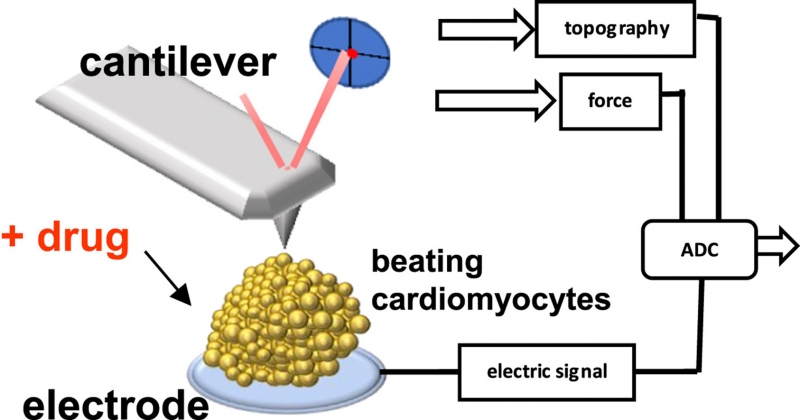
Cardiomyocytes (CM) placed on microelectrode array (MEA) were simultaneously probed with cantilever from atomic force microscope (AFM) system. This electric / nanomechanical combination in real time recorded beating force of the CMs cluster and the triggering electric events. Such "organ-on-a-chip" represents a tool for drug development and disease modeling. The human pluripotent stem cells included the WT embryonic line CCTL14 and the induced dystrophin deficient line reprogrammed from fibroblasts of a patient affected by Duchenne Muscular Dystrophy (DMD, complete loss of dystrophin expression). Both were differentiated to CMs and employed with the AFM/MEA platform for diseased CMs’drug response testing and DMD characterization.The dependence of cardiac parameters on extracellular Ca2+ was studied. The differential evaluation explainedthe observed effects despite variability of biological samples. The
β-adrenergic stimulation (isoproterenol) andantagonist trials (verapamil) addressed ionotropic and chronotropic cell line-dependent features. For the first time, a distinctive beating-force relation for DMD CMs was measured on the 3D cardiac in vitro model.


