ROLE OF CARDIAC MAGNETIC RESONANCE AND ECHOCARDIOGRAPHY PRIOR TO CARDIAC RESYNCHRONIZATION THERAPY
Introduction
Cardiac resynchronization therapy (CRT) is well established method for treatment of patients with heart failure. However, there are a substantial number of non-responders to CRT. A promising method appears to be an optimal left ventricular lead implantation based on modern imaging methods.
Methods:
27 patients underwent ECHO and cardiac magnetic resonance prior to CRT device implantation . The aim of the imaging studies was to determine an optimal pacing site for the left ventricular lead placement. The off-line analysis was performed by two experienced readers in a blinded fashion.
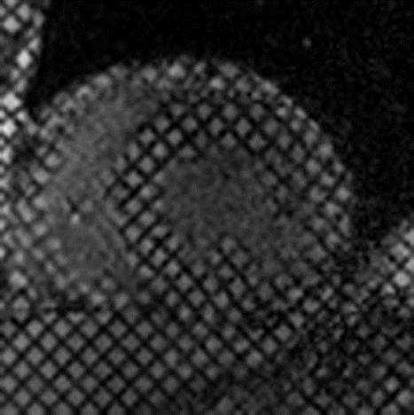
CMR was performed on 3T system. Cine, late gadolinium enhancement and myocardial tagging sequences were acquired. Image software Segment and inTag software were used for image analysis.
Standard echocardiographic parasternal and apical views were stored and analysed off-line. Left ventricular radial, longitudinal and circumferential strains were assessed using commercially available software.
Results:
Wall motion analysis was possible in all 459 segments in CMR and in 96% in echocardiography studies. Mean ejection fraction of the left ventricle was 29 % and 23% for ECHO and CMR. Scar transmurality was significantly overestimated by ECHO using the longitudinal strain cut-off -4.5% for discrimination between transmural and nontransmural scar.
The dyssynchrony analysis targeted to define the latest activated LV segment showed significant agreement between the two methods. The exact segment agreement was detected in 17 cases (63%). Discordance between target segment was detected in 4 patiens (15 %).
Summary and Conclusions:
We have demonstrated that CMR has a potential to improve diagnostic preoperative assessment in patients with severe heart failure and might be a useful tool for LV dyssynchrony assessment.
